Helmholtz Imaging Projects
Helmholtz Imaging Projects aim to initiate cross-cutting research collaborations and identify innovative research topics in the field of imaging and data science.
Funds for Helmholtz Imaging Projects are annually granted to cross-disciplinary research teams for collaborative mid-term projects.
Ideally, Helmholtz Imaging Projects are co-created with users and non-academic stakeholders to ensure the quick adoption of results.
Funding for the first Helmholtz Imaging projects started in December 2020. Many teams have since begun work on major challenges and pressing issues facing society to develop sustainable solutions for tomorrow and beyond.
Discover these outstanding and fascinating research projects with us or become a part of Helmholtz Imaging Projects and apply for your own project. The next call for Helmholtz Imaging Projects is OPEN until July 30, 2025. Find out more about the project call in this summary.
Helmholtz Imaging Projects – ongoing
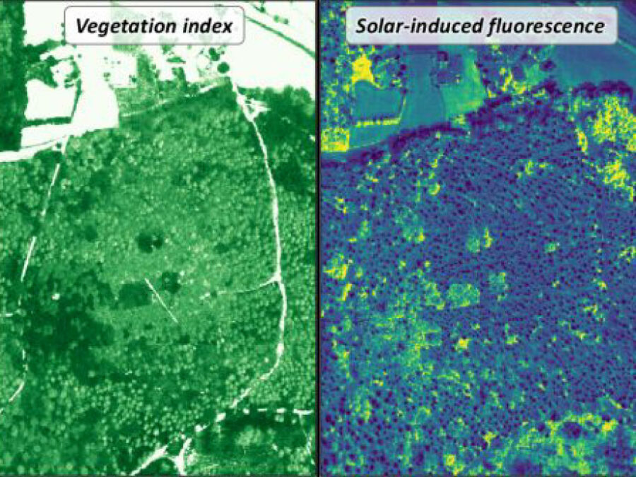
3DforestSIF
Understanding the solar-induced fluorescence (SIF) signal of natural, complex tree canopies
3DforestSIF seeks to correct airborne solar-induced fluorescence (SIF) data from forests for canopy structural and illumination effects, providing valuable insights for the early detection of forest stress.
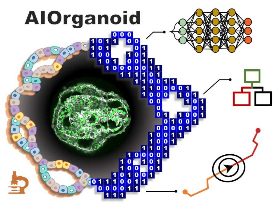
AIOrganoid
Artificial Intelligence Assisted-Imaging for Creating High-yield, High-fidelity Human Lung Organoid
AIOrganoid will apply cutting-edge imaging techniques and develop novel AI-based solutions to facilitate human lung organoid formation with high yield and fidelity, bridging the gap between cell biology and computational imaging.
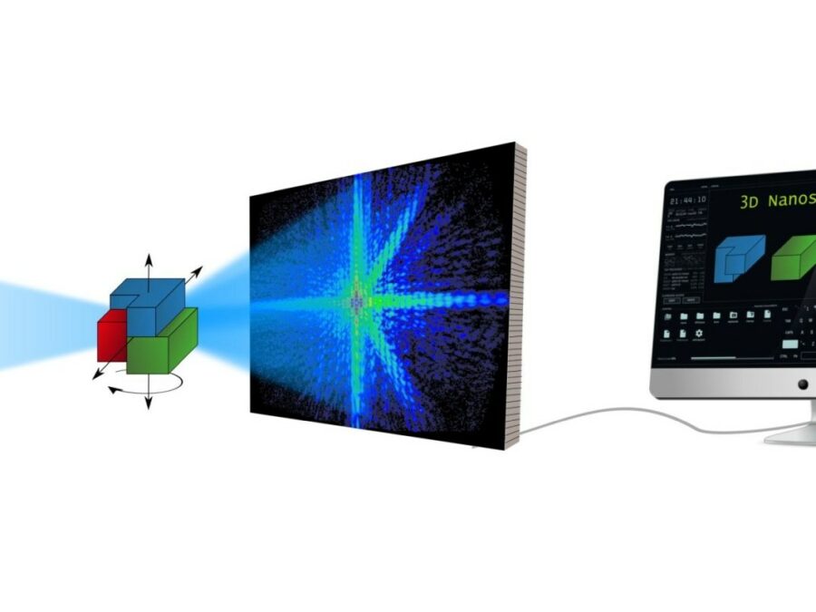
AsoftXm
Advanced Soft-X-Ray Microscopy Solutions
The project aims to develop a method that will speed up the analysis of diffraction patterns that arise in UV and soft X-ray light microscopy, so that the structure of the studied sample can be calculated more efficiently. The method could make the three-dimensional study of nanomaterials considerably easier. There are times when researchers need […]
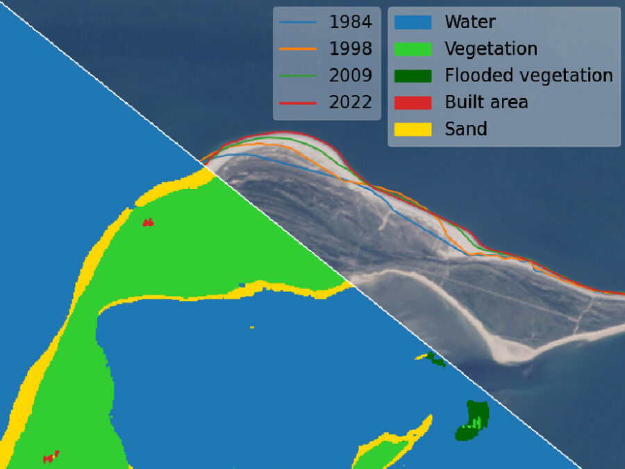
AutoCoast
Automatic detection of coastline change and causal linkage with natural and human drivers
Coastal erosion enhanced by climate change has become an increasing global threat, which requires rapid detection and reliable risk assessment. AutoCoast aims to provide advanced and reliable remote sensing-based AI tools to quantify coastline change rate at high-resolution and unravel the linkage between coastline change rate and natural and anthropogenic drivers at regional to global scale.
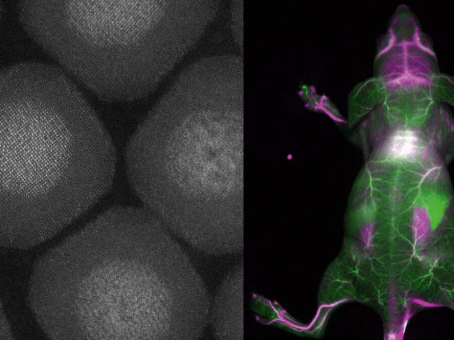
BENIGN
Biocompatible and Efficient Nanocrystals for Shortwave Infrared Imaging
The BENIGN project aims to enable non-invasive molecular imaging with cellular resolution in vivo at depths of several millimeters. This will be achieved using light from the shortwave infrared (SWIR) range (1000-2000 nm), which has less scattering and autofluorescence compared to the visible and near-infrared spectral range. Bright and targeted imaging agents are needed to fully exploit this range. The project will develop a new approach using lanthanide-based core-shell structures that emit light in the 1500-2000 nm range.
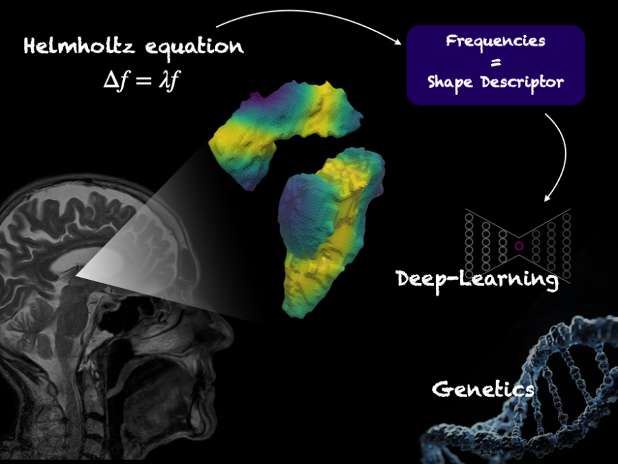
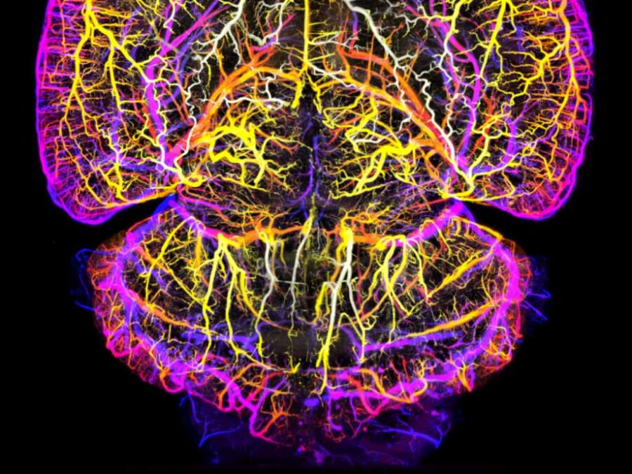
BrainShapes
Laplace-Beltrami shape descriptors of brain structures: Comparative optimization and genetic dissection
The project explores the 3D structure of the human brain by creating a digital ‘map’ of the brain and examining its unique genetic properties, potentially linking genetic variations to brain disorders.
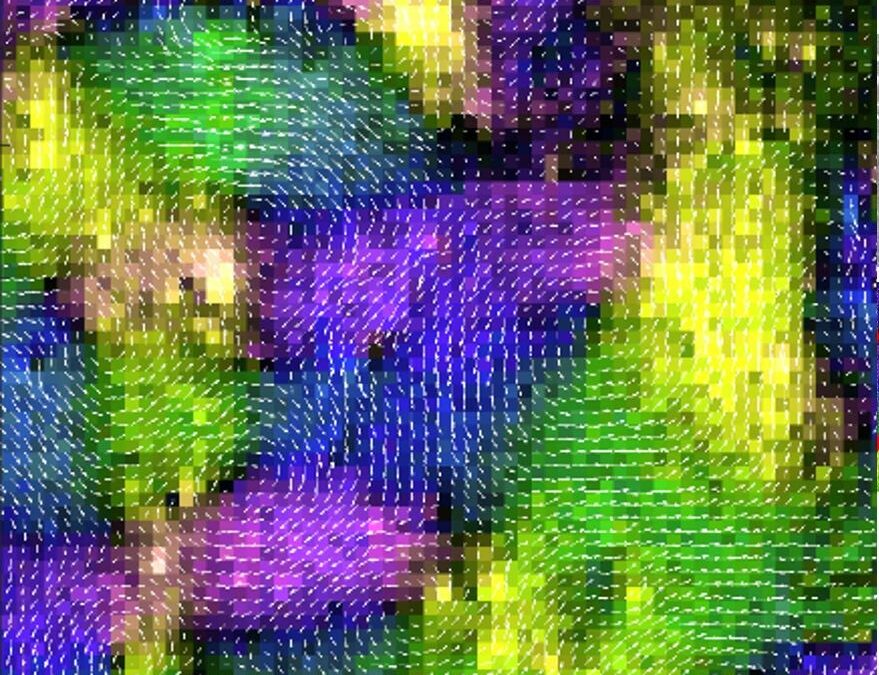
BRLEMM
Breaking resolution limit of electron microscopy for magnetic materials
A new method will make it possible to take images of the magnetic properties of materials under the electron microscope and to correlate these properties with their atomic structure. In order to achieve high resolution, a special algorithm must be developed to compute the magnetic properties from the microscope data.
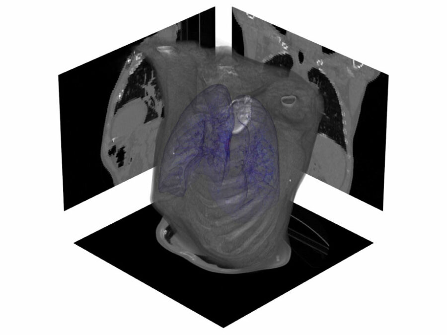
CLARITY
CineMR-guided ML-driven Breathing Models for Adaptive Radiotherapy
Dose-escalated radiotherapy of lung cancers requires precise monitoring of lesions and nearby organs at risk. Current methods are able to track ultra-central lesions but neglect their deforming vicinity, risking unacceptable toxicity to aortico-pulmonary structures. AI-based anomaly detection and generative AI models can address both requirements in real-time.
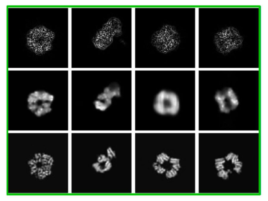
cryoFocal
3D Reconstruction from defocused cryo-EM images
This project explores how defocused images recorded with an electron microscope can be used to reconstruct the 3D structure of molecules inside cells. This method aims to enable faster and more cost-effective structural analysis of molecules to accelerate understanding of their functions and to design drugs against them.
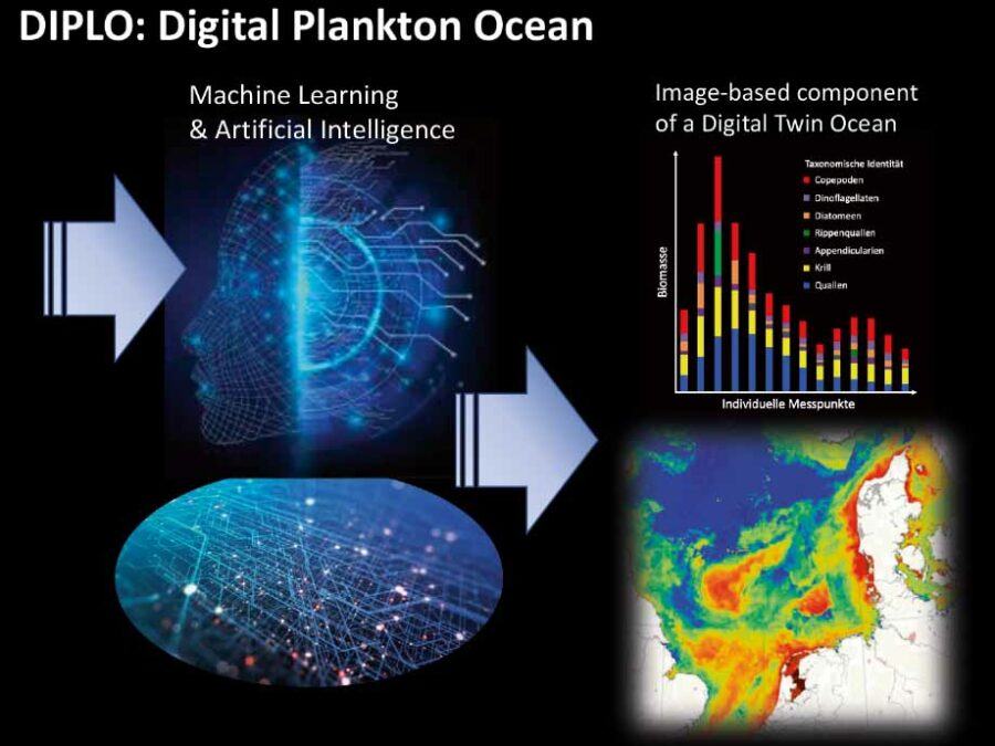
DIPLO
Paving the way from in situ plankton image data to a Digital Twin Ocean
This project will develop a user-friendly software platform to analyze plankton images independent of the instrument with which images were collected. This will help to compare data and create a common database, which is a critical step towards an image-based ecosystem component of a “Digital Twin Ocean”.
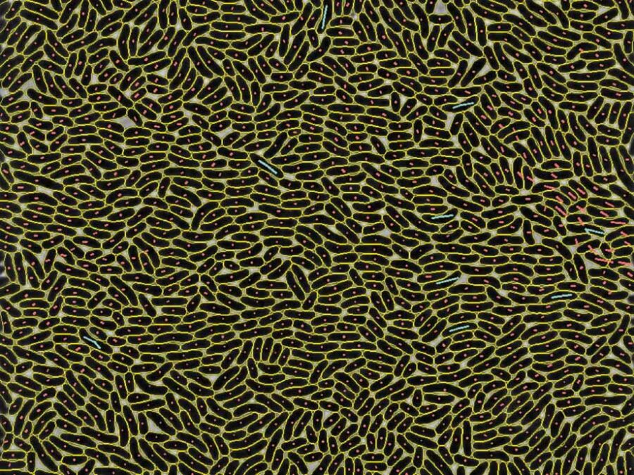
EMSIG
Event-driven Microscopy for Smart Microfluidic Single-cell Analysis
Microfluidic live-cell imaging (MLCI) unlocks spatio-temporal insights into population heterogeneity emerging from a single cell. EMSIG brings smart live-event detection capabilities to MLCI to facilitate the adaptive optimization of biological event resolution and autonomously counteracting deteriorating image qualities.
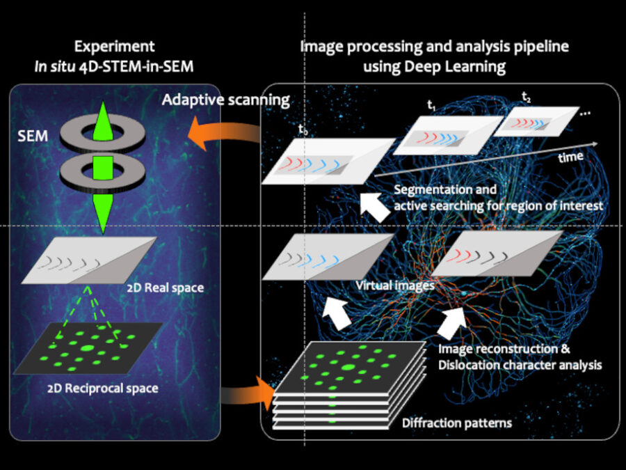
Fast-EMI
Deep-learning assisted fast in situ 4D electron microscope imaging
A novel imaging approach combining electron microscopy and deep learning has been established. This method enables adaptive tracking of atomic defects, accelerating material development for the energy transition.
FOMIA
A Foundation Model for Microscopy Image Analysis
This project will develop a foundation model trained on a large and diverse dataset of microscopy images to facilitate the adaptation of artificial intelligence methods to biological image analysis.
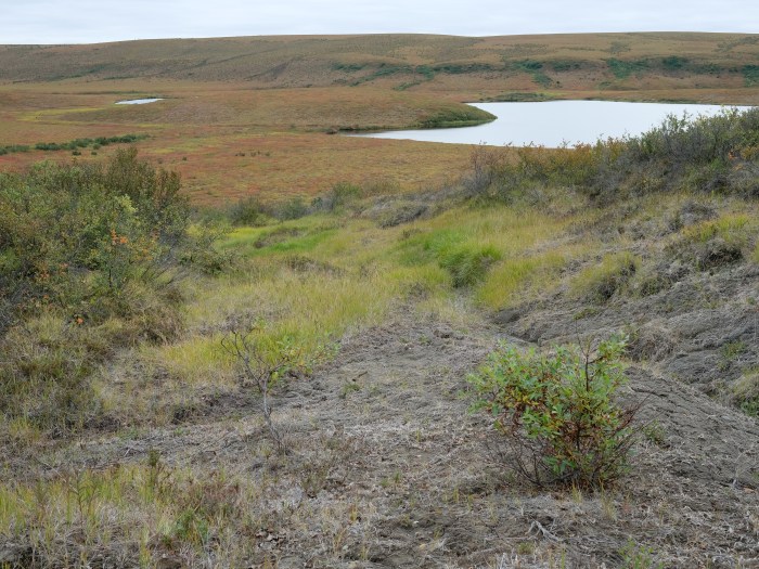
HIT Permafrost
The Hidden Image of Thawing Permafrost
The project aims to develop a method for determining just how extensively thaw processes have already progressed in permafrost regions. The machine learning approach to be developed will be used to analyse radar images from aircraft in order to learn more about the properties of the subsurface permafrost.
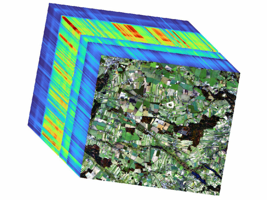
HYPER-AMPLIFAI
Advancing Visual Foundation Models for Multi-/Hyperspectral Image Analysis in Agriculture/Forestry
The project aims to make advanced AI models accessible for Hyperspectral Earth Observation, reducing computational demands, and improving environmental assessments through user-friendly interfaces.